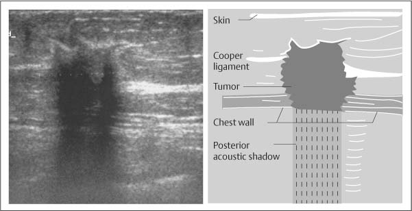
Posterior acoustic shadowing PAS can bias breast tumor segmentation and classification in ultrasound images. AbstractBreast ultrasound US in conjunction with digital mammography has come to be regarded as the gold standard for breast cancer diagnosis.
![]()
Transverse Ultrasound Of The Left Breast Demonstrates An Irregular Download Scientific Diagram
Breast sonogram reveals focus of intense acoustic attenuation without mass lesion.

. Ultrasound has become a useful adjunct modality for breast tumor diagnosis because it is cost-effective nonin-vasive and performed in real time 2. Which usually do not shadow. Newcomers to breast ultrasound may mistake the nipple for a breast mass because of its hypoechoic appearance shadowing and the intense vascularity beneath it.
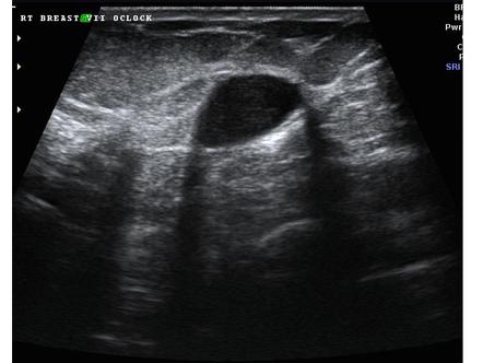
Posterior acoustic shadowing PAS can bias breast tumor segmentation and classification in ultrasound images. B Sonogram obtained after mild addi-. Hypoechoic irregular mass with posterior shadowing indistinct margins and vascular flow measuring 11 x 4 x 9 mm.
Anyone have benign results with posterior acoustic shadowing. Sonographic posterior acoustic shadowing. Variable sized fat deposits surrounded by foamy.
This loss is displayed in the image as shadowing and is an important sonographic sign for the detection and diagnosis of breast disease. While breast US has certain advantages over digital mammography it suffers from image artifacts such as posterior acoustic shadowing PAS presence of which often obfuscates lesion margins. Although posterior acoustic shadowing is a sonographic feature that is most commonly associated with mammary malignancies this sonographic finding may also be seen with benign breast lesions.
Posterior acoustic shadowing Non-circumscribed margins Non-palpable Controversial and age dependent Not new Case 3. Keywords Ultrasound Breast tumor Posterior acoustic shadowing PAS Half-contour feature Standard deviation of degree SDD 1 Introduction Breast cancer is a health problem for women worldwide 1. 70 F remote history of breast cancer and prior lumpectomy.
Acoustic shadowing orig-inates from Coopers ligament be-tween normal fat lobules. A Breast sonogram reveals focus of intense acoustic attenuation without mass lesion. You will be shown possible ultrasound correlates on the next slide and be asked to pick the best correlates and.
However knowledge of the shadowing vascularity and the ability to correlate the mass with the nipple on physical examination will help prevent newcomers from making this mistake. Upon a screening mammogram and ultrasound they found a 16 oval mass on my right breast. Acoustic shadowing from Coopers suspensory ligament mimics breast neoplasm at sonography of nor-mal breast in 43-year-old woman.
Volume 23 Issue 1. It is the posterior acoustic shadowing that is freaking me out. While breast US has certain advantages over digital mammography it suffers from image artifacts such as posterior acoustic shadowing PAS presence of which often obfuscates lesion margins.
It is wider than tall with macrolobulations no calcifications and posterior acoustic shadowing. The calculi in the kidney and gallbladder cause pronounced posterior acoustic shadowing. On mammography the lesion usually shows localized increased density in the glandular tissue.
The sonographic features of a benign solid mass include hypoechoic echotexture smooth circumscribed margins and lack of posterior acoustic shadowing 17. Breast ultrasound US in conjunction with digital mammography has come to be regarded as the gold standard for breast cancer diagnosis. An irregular hypoechoic mass with intense posterior acoustic shadowing can be typically seen on US and can mimic breast malignancy Fig.
Posterior acoustic shadowing is suspicious for breast cancer If a breast lesion shows posterior acoustic shadowing on ultrasound this means that there is something about the mass or around the mass which attenuates reduces the sonic beam strength in comparison to normal adjacent tissues. Fibroadenomas and lymph nodes are the most common benign solid masses encountered on screening breast ultrasound. Although posterior acoustic shadowing is a sonographic feature that is most commonly associated with mammary malignancies this sonographic finding may also be seen with benign breast lesions.
If a breast lesion shows posterior acoustic shadowing on ultrasound this means that there is something about the mass or around the mass which attenuates reduces the sonic beam strength in comparison to normal adjacent tissues. Retrospective analysis of 39 patients with posterior acoustic shadowing withoutassociatednoduleas a finding in breast ultrasoundthose lesions were subjected to ultrasound guided - tru-cut biopsy34 ortoexcisional biopsy 5withlocalization wire breastfrom January 2013 to April 2016Sonographicexaminationwas performed with 56-184 MHz. The phenomenon of acoustic shadowing sometimes somewhat tautologically called posterior acoustic shadowing on an ultrasound image is characterized by a signal void behind structures that strongly absorb or reflect ultrasonic waves.
It is a form of imaging artifact. Developing asymmetry upper inner breast mid depth. As ultrasonic beams propagate through tissues there is a loss of energy by absorption reflection and scattering.
The hepatic cyst demonstrates the opposite phenomenon of posterior acoustic enhancement. Acoustic shadowing originates from Coopers ligament between normal fat lobules. Multiple projections from the nodule within or around ducts extending away from the nipple usually seen in larger tumors.
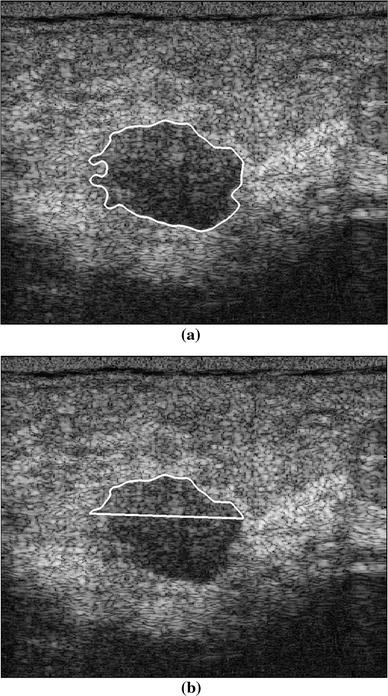
In this paper half-contour features are proposed to classify benign and malignant breast tumors with PAS considering the fact that the upper half of the tumor contour is less affected by PAS. Screen detected new mammographic mass Ultrasound is planned as the next step for this finding. Acoustic shadowing from Coopers suspensory ligament mimics breast neoplasm at sonography of normal breast in 43-year-old woman.
Shadowing may result because of reflection of most of the energy by a large impedance discontinuity. DMP usually shows nonspecific parenchymal enhancement rather than an irregular enhancing mass on MRI.

Ultrasound Image Of A Breast Cancer With Irregular Borders Angular Download Scientific Diagram
Sonographic Evaluation Of Benign And Malignant Breast Masses Iame

Posterior Acoustic Shadowing In Benign Breast Lesions Weinstein 2004 Journal Of Ultrasound In Medicine Wiley Online Library
Basic Principles Radiology Key
Benign And Malignant Characteristics Of Breast Lesions At Ultrasound Radiology Reference Article Radiopaedia Org

Posterior Acoustic Shadowing In Benign Breast Lesions Weinstein 2004 Journal Of Ultrasound In Medicine Wiley Online Library

Mediconotebook Posterior Acoustic Shadowing And Enhancement
Classification Of Benign And Malignant Breast Tumors In Ultrasound Images With Posterior Acoustic Shadowing Using Half Contour Features Springerlink
0 comments
Post a Comment